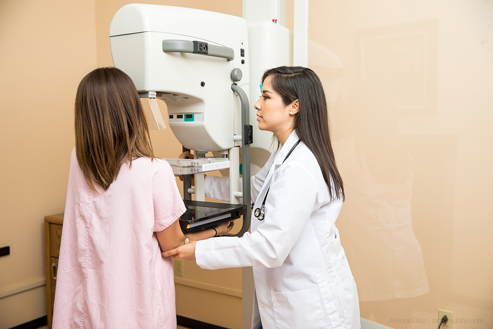
The objective of mammography is the radiological study of breast pathology. It has to be done from the age of 40 on an annual basis.
The problem with mammography is that it is uncomfortable for most women, and for a minority it can even be painful; everything depends on the pain threshold of each one, her predisposition and the fear of the final result.
Advantages of the Senographe Pristina system in mammography
Mammography is less bothersome when done within the first nine days after the start of menstruation. The main factor that favors them is the compression of the breast for a few seconds, with the aim of achieving a clear image. In this way, in addition, the gland is immobilized and the radiation to be used decreases.
However, with the new breast pressure self-monitoring system and the relaxation mechanisms that we use in our center, the discomfort is less.
This is Senographe Pristina, a technology that allows the patient herself to be able to control the compression of the breast, a fact that differentiates it from traditional mammography, which presses the chest automatically.
With this new tomosynthesis system, it is possible to obtain a very sharp and detailed image of the breast in 3D, with radiation exposure similar to that of a conventional mammogram and avoiding repetitions of the test (necessary when poor images are poor). . In this way, in some cases the patient receives less radiation, since she undergoes fewer tests.
Advantages of the Invenia ABUS system in mammography
Just as we did with Senographe, we have been the first to implement a comprehensive breast diagnosis system in Spain that includes automatic ultrasound equipment (Abus: Automatic Breast Ultrasound), capable of performing diagnostic tests in just 15 minutes.
Automatic breast ultrasound is an ultrasound technology that does not emit radiation and is especially indicated for testing dense breasts, that is, those in which there is a greater volume of breast tissue than fat. In this type of breast, up to a third of tumors can go unnoticed on traditional mammography because the breast tissue can mask the suspicious nodule, as it looks the same color on the image. Instead, using automated ultrasound, cancer is clearly differentiated from breast tissue.


